Caroline Stefani has a very cool job. She works at the Benaroya Institute in Seattle, USA, looking for new ways to treat diseases such as multiple sclerosis and type 1 diabetes. For this reason, Stefani spent a lot of time photographing the cells through a microscope and then conceived their three-dimensional shapes based on these photos.
This is not easy, and many scientists spend a lot of energy every day to think about the tiny three-dimensional structure of these cells or proteins.
Fortunately, Stefani's office was next door to Tom Skillman in the technical department. Skillman one day told Stefani that he had an idea to use microscope data to create a complete three-dimensional model of a cell in virtual reality.
A year later, Stefani's laboratories had a VR application in use that enabled her and other researchers to work in an unprecedented way. This is just one of many ways in which virtual reality and augmented reality can cause a stir in medical and medical research.
Adam Lacy-Hulbert, head of the laboratory where Stefani is based, said that this application is of great significance for the research work. Although it may seem a bit flashy, medical research mostly means trying repeatedly.
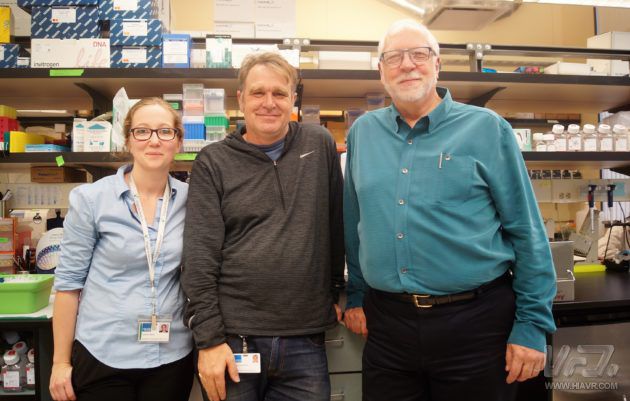
Caroline Stefani (left) and Adam Lacy-Hulbert (center) have been using VR to observe three-dimensional immune cells. This program was developed by Tom Skillman (right), the head of research technology.
Lacy-Hulbert's laboratory research on autoimmune diseases is the disease caused by the body's immune response to autoantigens and resulting in damage to their own tissues. Scientists spend a lot of time trying to get a list of what happened to cells, or how cells interact.
Lacy-Hulbert talked about the VR application he was putting into use: "It's cool, it's almost immediate!"
Technician Skillman said that seeing the Benaroya Institute scientists use this app is incredible. He said: "VR makes scientists very excited because they see something they have never seen before."
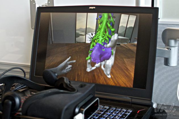
Briefly explain how the Confocal VR application works: First, the image is captured using a confocal microscope. The resulting image is a two-dimensional image of a cell taken from different angles, similar to the MRI in the hospital. .
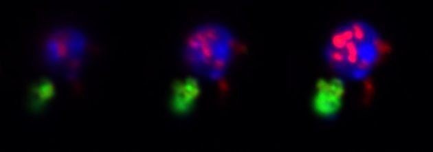
This is an image taken by a confocal microscope. Green is a dead cell that is swallowed by immune cells. The immune cell nucleus is blue.
Although these images help us to understand roughly where the cells are and how they interact, it is difficult to have a more real sense and it is difficult to see what actually happened. Even synthetic 3D images are difficult to understand thoroughly through computer monitors.
These images are then transferred to Skillman's Confocal VR program built using the popular video game creation software Unity. This process is very fast, Lacy-Hulbert said, basically when you go from the microscope to the VR head position .
He said: "You can quickly view these images under a microscope, and then you can study them in VR for the first time."
In addition to Skillman and Stefani, this project team has another researcher, Mridu Acharya. They use the rapid prototyping technology in the field of technology.
"I'll start by creating something for Stefani. She'll give me feedback after using it. Then I go update, adjust it, and iterate as fast as I can," Skillman said.
The final product is quite amazing: The program not only shows the cells to scientists in a new way, but it is also very quick to use.
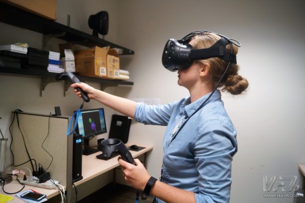
Personally, I have never used confocal microscopy before, nor have I studied the immune system in depth. When Stefani explained the details of the 2D image, I could hardly understand her. But once I put on the lab's Vive headline, I immediately understood the details she described. I can see more and ask the researchers about different parts of the cell.
This application also allows scientists to control and manipulate images. Confocal's VR Lab includes a control panel with sliders for adjusting brightness, contrast and lighting that are intuitive to use.
The intuitive nature of virtual reality is one of its strengths. Especially in the medical field, people constantly try to understand highly complex objects.
This technology is also attracting attention in hospitals and other medical institutions. One example is Seattle-based startup Pear Med, which is developing a hybrid reality program that can create 3D interaction models from patient scans, effectively showing patients and doctors anatomical 3D images of their organs and other parts.
Pear Med's CMO Seslar said the goal of the tool is to build better patient anatomy models in medical environments such as operating theatres through better visualization and 3D interactions. Giving patients a better understanding of their health status also helps doctors prepare for complicated operations.
Apps by Pear Med
Seslar said: "We expect that soon one day, our technology will be used to display the patient's anatomy in real time during the clinical course.
On the other hand, Benaroya is also developing a VR program that allows scientists to interact with protein structures in VR. These highly complex molecules are responsible for most of the cell's functions.
The application, called AltPDB, allows multiple people to collaborate in VR and is open to the public: anyone with a VR heads up and AltSpace account can try it in their own living room. They also plan to share Confocal VR with non-profit organizations.
Whether it's tiny proteins, small cells or whole organs, the mixed reality can help us see things in new ways.
Lacy-Hulbert said that scientists often see things that have never before been noticed through Confocal VR, although they have been studying this type of cell for decades. The immune cells engulf the image of dead cells is a good example.
You can see in the two-dimensional image that the cytoplasm seems to pass through its nucleus. Scientists can see some strange things happening in these images, but they are not sure what they are.
In VR, you can grab it, turn it to the right angle, and zoom in on it. "Lacy-Hulbert said.
He also said: "This is very important, because when people used to study this issue in the past, scientists thought that some proteins actually invaded the cell nucleus. Now we can tell them that they are two different structures, It's one that is pushing another."
In some cases, this detail may be a small change. But under certain circumstances, this may mean a new way to treat allergies, a development of emergency medicines, or other medical advances.
This article is original by VR net, please indicate VR net and chain back.
Small Air Winch,Air Raking Winch,Air Pneumatic Winch,Pneumatic Raking Winch
RUDONG HONGXIN MACHINERY CO.,LTD , https://www.rdhxmfr.com